,

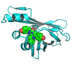
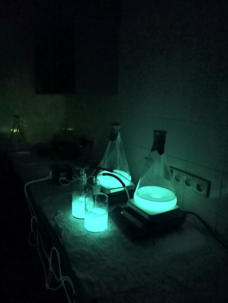
SCIENTISTS FROM BRNO HAVE REVEALED THE STRUCTURE OF THE BRIGHTEST ENZYME. THEY CAN MAKE IT GLOW EVEN MORE INTENSELY AND INCORPORATE IT INTO HUMAN CELLS

Bioluminescence, the ability to produce visible light by living organisms, is one of the most beautiful biological phenomena that has fascinated humanity for centuries. While there are not many terrestrial bioluminescent creatures, the situation is entirely different in the seas and oceans. In the dark depths of the oceans, various organisms literally abound, producing light in various colors. This ability is granted to them by luciferases, enzymes that endow living beings with the capability to emit light. However, how marine luciferases function has been a mystery until recently. Now, researchers from Masaryk University in Brno and the International Clinical Research Center (ICRC) have successfully uncovered the structural essence of this enzyme.

Shrimp luciferase is the brightest. And now we know how it works.

Scientists worldwide have been attempting to uncover the essence of this enzyme for decades, but it was only recently that researchers from Brno succeeded. They isolated one of the smallest yet brightest enzyme celebrities, called nanoluciferase, from the deep-sea shrimp Oplophorus gracilirostris.

When a predatory fish develops a taste for this shrimp, it, as a defensive mechanism, releases a highly viscous secretion into its surroundings, which soon transforms into a stunning light show. While the predator is baffled by this underwater fireworks display, the shrimp gains valuable time to escape.

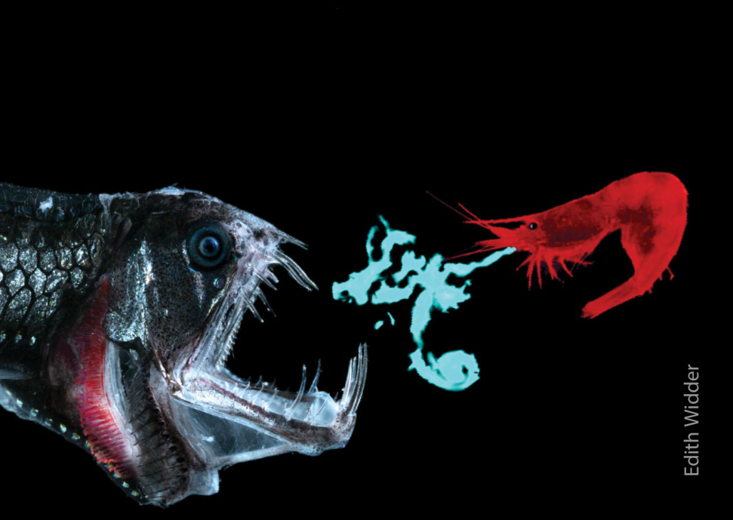
Shrimp defends against dragonfish

Oceanographer Edith Widder captured a deep-sea shrimp defending itself against a predator through bioluminescence.

Due to its truly small size, nanoluciferase enzyme is highly popular, and scientists and engineers worldwide use it for non-invasive imaging of various biological processes. In recent years, luciferases have also found applications in the photodynamic therapy of cancerous diseases.

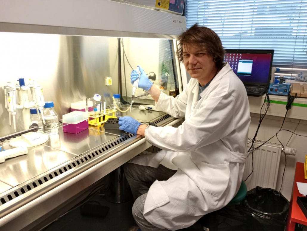
“However, despite the widespread popularity of nanoluciferase, the molecular mechanism by which this brightest luciferase produces light has not been known until now,” explains the head of the research team, Martin Marek, from the Loschmidt Laboratories of the Faculty of Science at Masaryk University and the International Clinical Research Center of St. Anne’s University Hospital and the Faculty of Medicine of Masaryk University.

Team Leader of Molecular and Structural Biology at Masaryk Univerzity Martin Marek.

Thanks to modification, the enzyme can glow even more intensely.

“In the laboratory, we successfully biochemically prepared nanoluciferase in complex with luciferin molecules and revealed the atomic structures of these macromolecular complexes. This allowed us to visualize the key steps of the nanoluciferase reaction and understand its reaction mechanism at the molecular level, that is, the essence of how nanoluciferase glows. As a result, we were able to make structural changes in the nanoluciferase molecule that led to its catalytic improvement,” added members of the research team, Jana Horáčková, and Daniel Pluskal. Thanks to the work of Brno scientists, this tiny enzyme can now glow even more intensely. The research team published their findings in the journal Nature Communications.

Researchers Jana Horáčková and Daniel Pluskal.

Glowing human cells: Advancements in cancer research and new drugs

The method for measuring and evaluating the efficiency of the modified luciferase was devised by Tomáš Bárta, the head of the Stem Cell Biology and Vision Research group at the Faculty of Medicine, Masaryk University. “In collaboration with Martin Marek, we obtained the sequence of the modified gene for nanoluciferase, and we incorporated this sequence into human cells. Using the device we developed, we thoroughly examined the activity of this modified luciferase. The results showed us that the modified form is much more active,” explains Bárta.

Head of the research group Tomáš Bárta with the device for measuring enzyme activity.

The discovery is a significant milestone. It opens up new possibilities for utilization across a wide spectrum of applications, from basic research to practical applications. The acquired knowledge will contribute to a better understanding of bioluminescence mechanisms and has the potential for the development of more sensitive bioluminescent reporters. This breakthrough will propel further research in human cells, testing new drugs, and the insights gained will find applications in cancer biology and the study of gene expression.

“We compared genetic modifications, searching for the one that most efficiently converts luciferin into light, meaning it will produce the most light from a given amount of substrate,” he adds.

However, the application of nanoluciferase is much broader. “Nanoluciferase is used, for example, in non-invasive monitoring of individual cells or viruses. Thanks to it, we can observe how metastatic cells spread in the organism or how a viral infection permeates it. Biological processes can thus be monitored in both time and space,” Marek adds.

Nanoluciferase in detail.

A cheap and accessible solution for the rest of the scientific world

And how to measure the activity of luciferin in a cell is not kept secret by the researchers. The research team led by Tomáš Bárta has introduced an open-source platform for constructing a universal, inexpensive, lightweight, and portable luminometer – LuminoCell, which can be 3D printed.

LuminoCell is capable of simultaneously measuring luciferase signals in six Petri dishes. And the process is not expensive at all. The estimated costs for assembling LuminoCell are approximately 40 US dollars, or a little over 800 Czech crowns. “It takes about an hour to assemble,” adds Bárta. It can be used in various types of cell culture incubators and is capable of performing sensitive real-time detections.